LATERAL ANKLE PROJECTION
Mediolateral View • Sagittal evaluation of the ankle joint and hindfoot
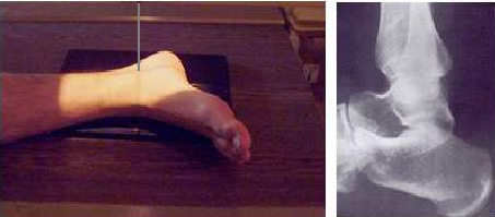
Proyección Lateral de Tobillo - Vista anatómica
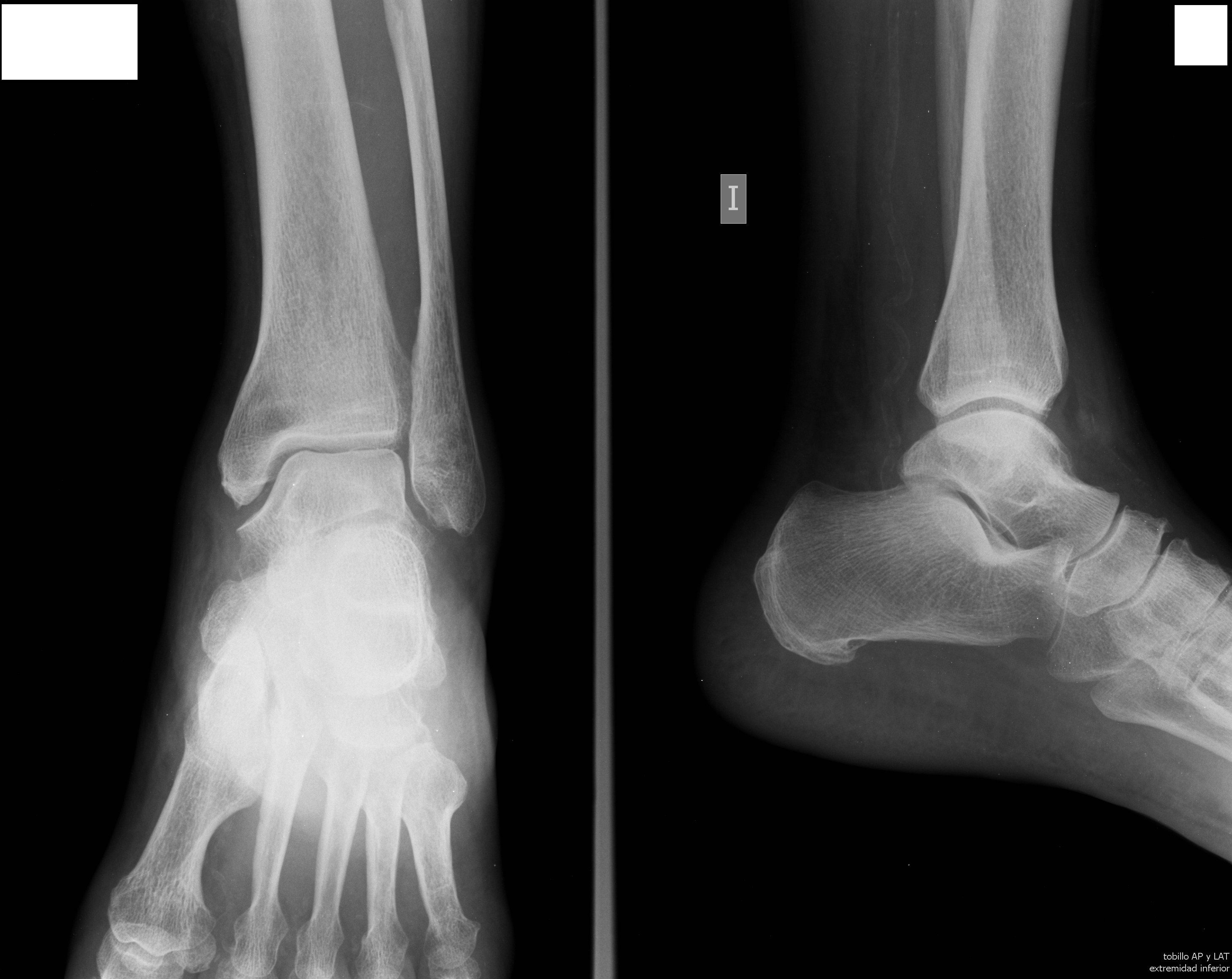
Posicionamiento Ap y Lateral de Tobillo - Vista técnica
Exposure Factors
50-60
Kilovoltage (kV)
6-10
Milliampere-seconds (mAs)
Fine/Broad
Focal Spot Type
100-110 cm
Source-Image Distance
Equipment: Generally without bucky (tabletop). Position: Lateral decubitus.
Cassette Size
24 × 30 cm
Crosswise (Transversely)
Visible Anatomical Structures
Distal Leg
Fibula and Tibia (distal third)
Tibiotalar Joint
Profile of the joint space
Tarsal Bones
Talus, Calcaneus, Cuboid, Navicular
Metatarsals
Base of the 5th metatarsal
- Distal Fibula superimposed over the posterior half of the Tibia
- Medial and Lateral Malleoli superimposed
- Tibiotalar Joint clearly visible in profile
- Talus and Calcaneus in lateral profile
- Subtalar Joint (Articulatio subtalaris)
- Navicular and Cuboid bones
- Base of the 5th metatarsal (important for Jones fracture)
- Pre-achilles and Pre-patellar fat pads (signs of joint effusion)
Patient Positioning
Patient in lateral decubitus on the side of the affected limb
Affected limb flexed at the knee for stability
Unaffected limb moved away (posteriorly or anteriorly) to avoid overlap
Rotate the limb until a true lateral position of the foot and ankle is achieved
Dorsiflex the foot to 90° with the leg (if the patient's condition allows)
Malleoli should be aligned vertically (perpendicular to the cassette)
Center the ankle in the middle of the selected portion of the cassette
CENTERING POINT
Medial Malleolus
The central ray is directed to the medial malleolus of the ankle
Central Ray
Direction: Vertical and perpendicular to the long axis of the leg and the plane of the cassette.
Angulation: 0° (Perpendicular).
Image Quality Criteria
- Complete visualization of the distal tibia and fibula, talus, and calcaneus.
- True lateral position: the distal fibula is superimposed over the posterior half of the tibia.
- The tibiotalar joint space should be open and centered in the image.
- Overlap of the medial and lateral malleoli.
- Visualization of the base of the 5th metatarsal and the tuberosity of the calcaneus.
- Optimal density and contrast to see both bone structures and soft tissues (fat pads).
Clinical Indications
Malleolar fractures
Ligamentous injuries
Joint effusion
Foreign bodies
Radiological Tip
Optimization for distal extremity studies:
- 24 × 30 cm divided transversely in half
- Resource optimization - Two studies using one cassette
- Cost reduction - Efficiency in radiographic material
- Appropriate size - Sufficient for the anatomical region of the ankle
This practice is especially useful in emergency services with high patient volume